Unlocking the Power of Medical Images for Surgeons
Unlocking the Power of Medical Images for Surgeons
Kidney Aneurysm: See inside the aorta and the renal artery to visualize a kidney aneurysm inside and out
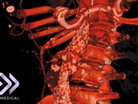
Aortic Dissection: Immersively visualize an aortic dissection including entry points, true and false lumen, fenestrations and dissection flap with your patient CT scan
Aortic Arch Aneurysm: Enter the aorta to visualize the aortic arch aneurysm of a patient with congenital heart disease

Endovascular vs Vascular
Access Site and Implant Selection
Implant Placement Confirmation